Failure of the molecular bodyguard in Parkinson’s disease
Scientists from ETH Zurich and the University of Basel’s Biozentrum have shown that chaperone proteins dynamically bind to the Parkinson protein α-synuclein. If this interaction is disturbed, it leads to cell damage and the formation of aggregates typical for the disease.
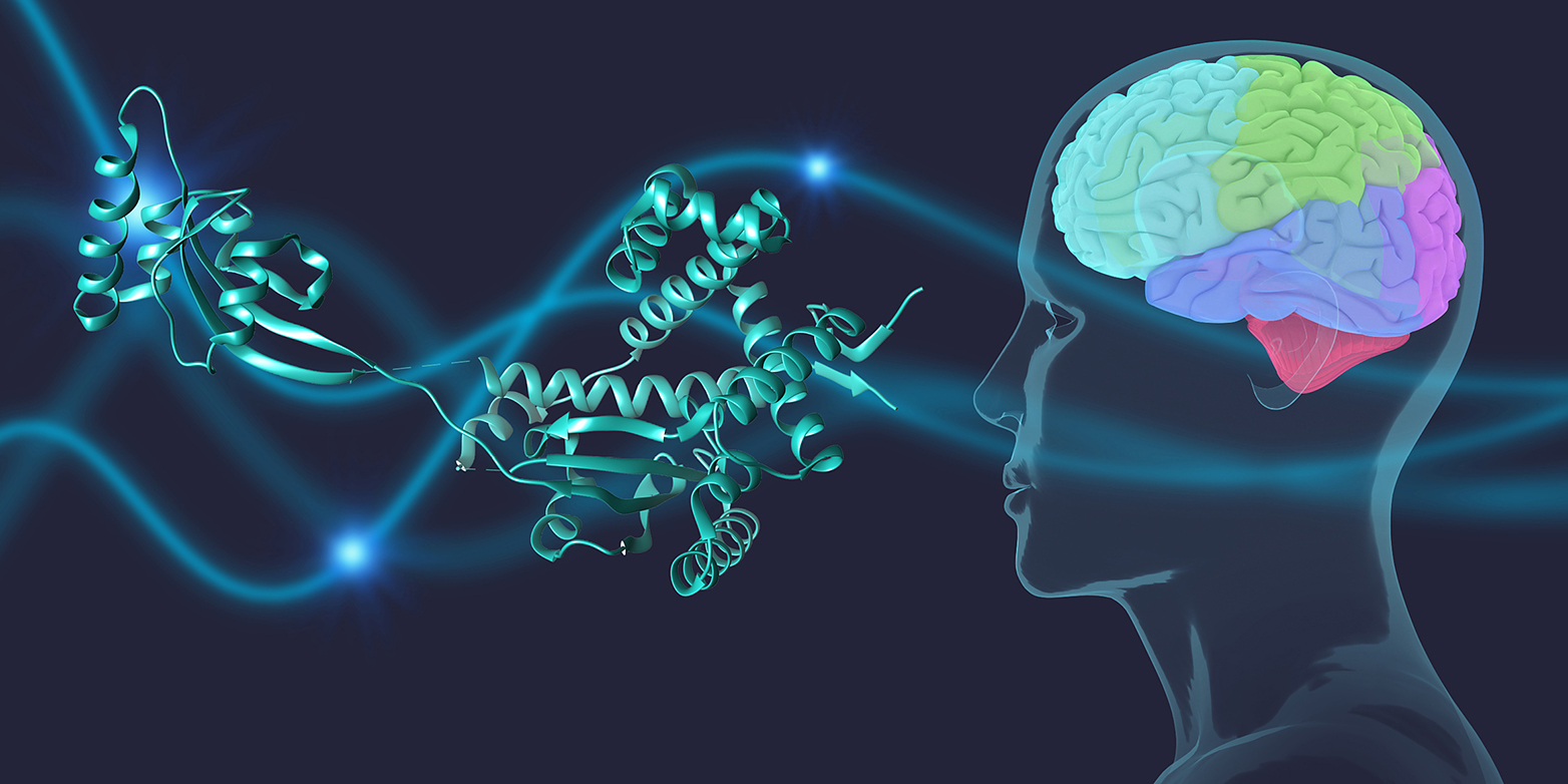
Parkinson’s disease is characterised by the progressive death of nerve cells in the brain. Its precise causes are still unknown, which is why it’s so difficult to develop effective therapies. It is known, however, that α-synuclein, a protein found in the brain, plays a role in this disease.
Researchers at ETH Zurich and the University of Basel’s Biozentrum have now discovered that chaperone proteins are also involved in the development of Parkinson’s: in healthy cells, α-synuclein is always surrounded by these chaperones, which ensure that α-synuclein remains functional. But when the chaperones can no longer fulfill their bodyguard function, this can have grave implications.
For example, chemical modifications of α-synuclein – as observed in Parkinson’s – can disturb its interaction with the chaperones, or the chaperones themselves can become less active with age. One result of this is that “unaccompanied” α-synuclein proteins bind to cellular membranes, such as those of the mitochondria (the power plants of the cell), where they aggregate and concomitantly at least in part gradually destroy the membranes. The membrane debris and α-synuclein then combine to form Lewy bodies, as other scientists have recently shown. These intra-cellular aggregates are typical of Parkinson’s.
Scientists identify new chaperone role
Chaperones were previously known for helping proteins to retain their functional 3D structure. When proteins are built in the cells they are in a chainlike form. Chaperones bind to these and therewith ensure that they fold to the proper 3D structure. “In our work, we’re now showing that chaperones have an additional job: by dynamically binding to the proteins, they can also regulate their function,” says Roland Riek, Professor of Physical Chemistry at ETH Zurich. He led the project together with Sebastian Hiller, Professor at the University of Basel’s Biozentrum. Their paper is published in the journal external page Nature.
The researchers were able to show that chaperones dynamically bind to α-synuclein using in-cell NMR spectroscopy. NMR spectroscopy allows scientists to explore the 3D structure of molecules and macromolecules. Normally, this method requires purified molecules in solution. The relatively new in-cell NMR spectroscopy method, however, enables measurements to be taken directly inside biological cells.
The new findings could provide fresh impetus to efforts in treating Parkinson’s. According to the scientists, chaperones and the maintenance of their function should also be considered in the development of novel therapies.
This article is based on a external page text by the Biozentrum of the University of Basel.
Reference
Burmann BM, Gerez JA, Matečko-Burmann I, Campioni S, Kumari P, Ghosh D, Mazur A, Aspholm EE, Šulskis D, Wawrzyniuk M, Bock T, Schmidt A, Rüdiger SGD, Riek R, Hiller S: Regulation of α-synuclein by chaperones in mammalian cells. Nature, 4 Dezember 2019, doi: external page 10.1038/s41586-019-1808-9
Comments
No comments yet