A new direction for cancer research
In collaboration with University Hospital Basel, researchers from ETH are investigating the early stages of bladder cancer. Their findings show that future research should also focus on mechanical changes in tumour tissue.
Dagmar Iber is Professor of Computational Biology at ETH’s Department of Biosystems Science and Engineering in Basel. Her research group uses a combination of lab experiments and computer modelling to investigate how cells organise themselves into organs and other complex, three-dimensional tissue structures based on the genetic information they contain. Until recently, their work did not touch on cancer research. But that all changed when the ETH Board issued a call for research proposals combining basic and medical research on new topics in health-related fields.
In response, Iber teamed up with two professors from the University Hospital Basel, urologist Cyrill Rentsch and pathologist Lukas Bubendorf. They were seeking to understand what governs the direction in which bladder tumours grow. As it turns out, their collaboration may well have provided cancer research with a major new lead.
Tumour type is key
The direction in which a bladder tumour grows can play a key role in whether it proves malignant or benign. In turn, this also determines the course of treatment and the patient’s chances of survival.
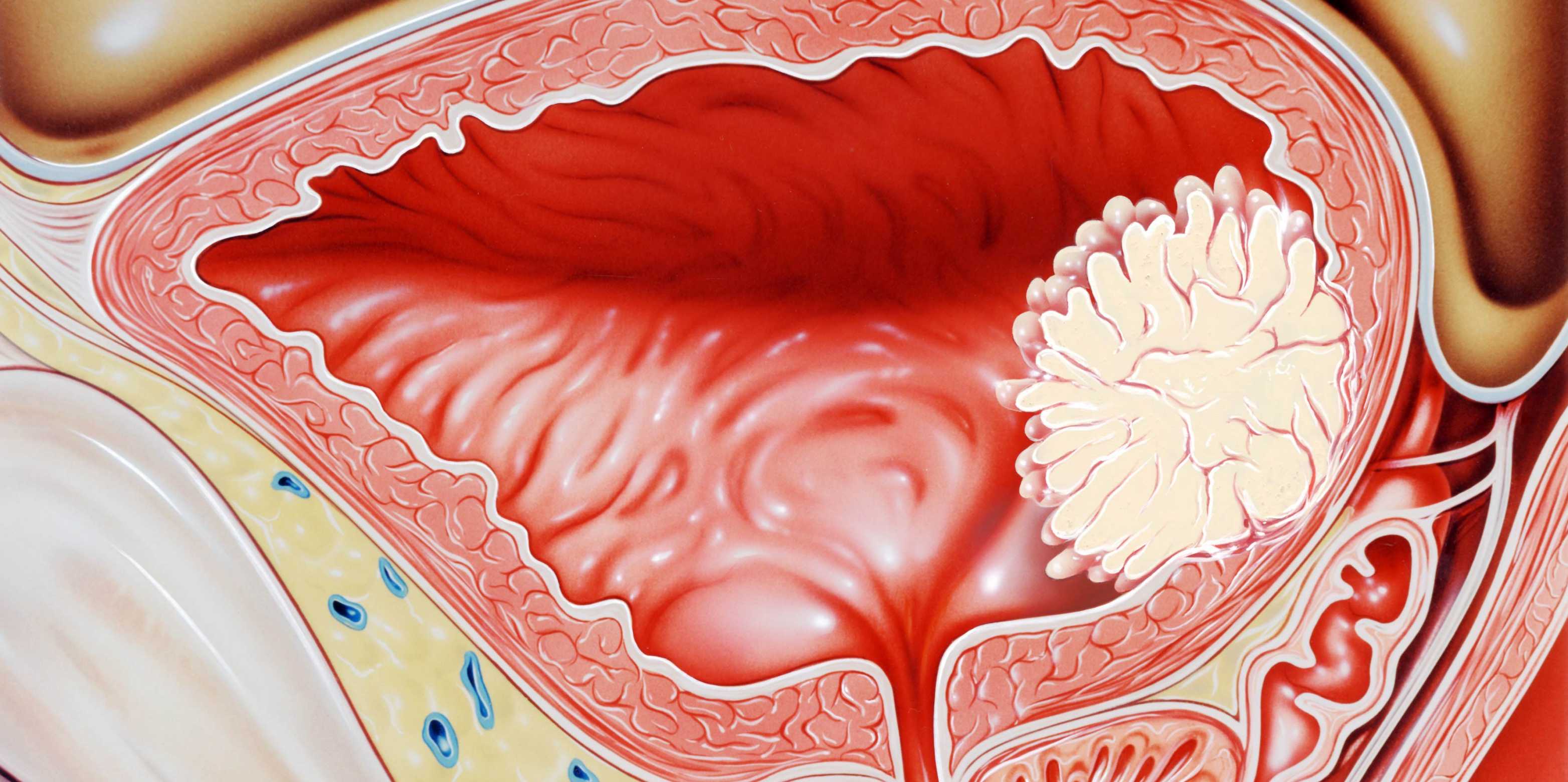
One of the most common forms of bladder cancer is a papillary tumour. This has a slender, branch-like structure and grows from the bladder wall into the bladder cavity. It is a relatively benign form of bladder cancer that can be effectively treated in a minimally invasive procedure that involves scraping the tumour off the bladder wall.
Harder to treat is what doctors refer to as muscle-invasive bladder cancer, where the tumour grows not into the bladder cavity but rather into the deeper layers of the bladder wall. Access to blood and lymphatic vessels in those deeper layers facilitates the formation of metastases that can then spread through the body. In this case, the prognosis is much less favourable; frequently, the entire bladder must be surgically removed. It is known that these two forms of cancer differ genetically. However, these differences do not explain why one cancer type would grow into the inner layers of the bladder wall and the other grow out into the bladder cavity.
About
Dagmar Iber is Professor of Computational Biology at the Department of Biosystems Science and Engineering and President of the University Assembly.
The team’s initial inspiration came from their work on lung development. “The tree-like ramification of the papillary bladder tumours have some likeness to the minute branches of the bronchioles in the lungs,” Iber explains. This led them to wonder whether similar molecular mechanisms might be responsible for creating both these structures. Yet subsequent research showed this not to be the case. “It turns out that the molecular drivers in the formation of lung tissue are quite different to those in the development of bladder cancer,” says Iber.
Mechanical, not biochemical
When it comes to lung tissue, a biochemical mechanism defines the position of new branches. In bladder cancer, however, the emergence of papilla appears to be influenced by mechanical rather than biochemical factors. In support of this theory, the Basel-based researchers have now published a study in the form of a preprint – a full draft of a research paper that is shared publicly before peer review.
To understand their theory, it helps to visualise the structure of the bladder wall. This wall is flexible and has numerous folds that enable the bladder to expand and contract depending on the amount of urine it has to hold. Three layers of tissue play an important role here. Together, they make up the innermost layers of the bladder wall, like an onion: first, a soft epithelial layer on the inside of the bladder wall; next, a substantially stiffer membrane, which provides mechanical stability; and, beyond that, a somewhat softer layer of connective tissue.
Based on computer models, biopsies from tumour patients and tissue samples harvested from experiments with mice, the researchers’ theory posits that cancerous growth is accompanied by changes in the relative stiffness of the different layers of the bladder wall. Depending on the degree of these changes, different forms of cancer develop. If there is only a minimal change in the stiffness of the layers relative to one another, blunt protrusions may grow from the bladder wall into the cavity of the bladder. These can then form the basis for papillary tumours. By contrast, if changes in relative stiffness are more significant, the surface of the bladder mucosa remains smooth. Instead, the membrane that separates the epithelium from the surrounding layer of connective tissue forms fine wrinkles and narrow folds. The researchers posit that this can result in tissue damage that encourages the growth of a malignant tumour into the inner layers of the bladder wall.
Focus on the early stages
“Pathologists have described changes in bladder wall stiffness in patients with advanced bladder cancer,” explains Franziska Lampart, a doctoral student in Iber’s group. The Basel researchers now used an animal model to investigate the early stage of bladder cancer. In collaboration with the group led by Daniel Müller, Professor of Biophysics at the ETH Department of Biosystems Science and Engineering, atomic force microscopy revealed localised softening of the membrane layer even at this early stage. “This supports our idea that local changes in the relative stiffness of individual bladder wall layers play an important role in the development of bladder cancer,” says Lampart.

“Cancer research needs to focus more closely on biomechanics and the chemical signalling pathways that affect it.”Dagmar Iber
These findings may well take cancer research in a new direction. At present, much of this work focuses on inhibiting the growth of cancer cells or killing them. “But our research shows once again that tissue mechanics is important, too,” Iber notes. Cells secrete protein fibres and enzymes that influence and modify the extracellular matrix that surrounds them. “Cancer research needs to focus more closely on biomechanics and the chemical signalling pathways that affect it,” Iber says. “But this line of investigation is still very much in its infancy.”
Besides the enjoyment of constructive collaboration with clinicians from University Hospital Basel, this cancer research project has brought Iber new insights. These may well prove invaluable back on her home territory of developmental biology, where tissue mechanics also play a role.
Globe The Basel connection

This text appeared in the 24/01 issue of the ETH magazine Globe.
Download Read whole issue (PDF, 3.5 MB)


Comments
No comments yet