Faster diagnosis of endometriosis with AI
ETH spin-off Scanvio is developing an algorithm that can be used to analyse ultrasound images of the womb on an automated basis. This should enable doctors to diagnose endometriosis more quickly in the future.
In brief
- Around ten percent of all women of childbearing age are affected by endometriosis. The growths of the endometrium regularly cause severe pain during menstruation.
- ETH spin-off Scanvio has developed an algorithm intended to assist gynaecologists in better interpreting ultrasound data of the womb in order to enable the early diagnosis of endometriosis.
- Endometriosis is at present largely diagnosed by means of laparoscopy. The algorithm from Scanvio is intended to enable this onerous and expensive procedure to be avoided much more in the future.
Endometriosis is widespread. Around ten percent of all women of childbearing age throughout the world suffer from it. And “suffer” is the operative word here, as it takes an average of eight to twelve years for these benign growths of the endometrium in the abdominal cavity to be diagnosed. Years in which women endure severe pain generally before and during menstruation.
In order to reliably diagnose endometriosis, many gynaecologists still rely on a laparoscopy performed under general anaesthetic. However, this procedure is not only laborious and onerous for patients but also relatively costly. While endometriosis could be diagnosed for most patients via ultrasound, this calls for a certain degree of experience as it can easily go undetected.
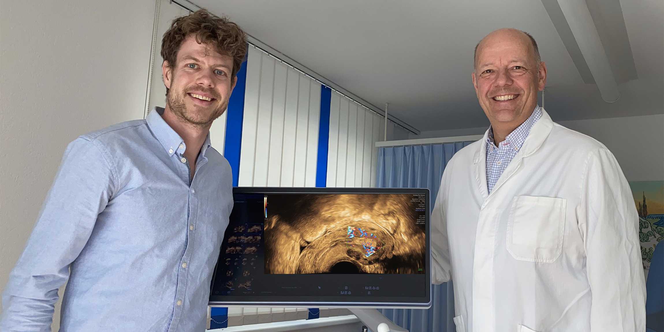
AI expert Fabian Laumer and gynaecologist Michael Bajka therefore founded the spin-off Scanvio in the summer of 2023. Their goal is to develop an algorithm to assist doctors in interpreting the ultrasound data of the womb during the initial examination in order to diagnose endometriosis reliable and much faster. They are receiving specialist support from the ETH AI Center and their two co-founders ETH Computer Science Professor Joachim Buhmann and Julian Metzler, an endometriosis specialist at University Hospital Zurich.
Entrepreneurs by chance
The fact that Laumer is today developing medical solutions is something he owes on two counts to chance. For although medicine and biology already fascinated him as a child, he initially studied electrical engineering and information technology. It was only when he was studying for his Master’s degree that he had the opportunity to combine artificial intelligence (AI) and medicine. “I heard by chance that Buhmann’s research group was offering a Master’s thesis on the AI-based analysis of ultrasound data of the heart,” explains Laumer. He immediately applied – and was successful.
After completing his Master’s degree he continued the research work in a doctoral project. And once again chance came to his aid. Bajka contacted his research group asking whether AI could be used to detect endometriosis. Laumer was exactly the right person for the gynaecologist specialising in endometriosis to put his question to. For his doctoral project, the ETH researcher developed an algorithm enabling the better interpretation of ultrasound data of the heart. This approach was then transferred to the womb.
Creating a 3D model from 2D images
Laumer and Bajka developed an algorithm that identifies pathologies on the ultrasound images of the womb that are often difficult or even impossible for the human eye to see. To this end, Laumer trained the algorithm with ultrasound images and patient data. “The number of pregnancies and Caesareans, age or phase in the menstrual cycle – all these factors obviously influence the appearance of the womb,” he explains.
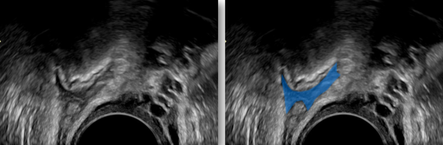
The algorithm currently shows the endometriosis in colour on 2D ultrasound images. If the development work proceeds as planned, Laumer hopes to generate a 3D model of the womb by the end of the year on which all growths and adhesions are clearly marked. This could enable gynaecologists to precisely localise endometriosis and better assess the severity of the condition.
Standards for endometriosis diagnosis
To ensure that the Scanvio AI solution delivers the most reliable results possible, Bajka and Laumer also wish to define fixed standards for the ultrasound examination of endometriosis. A software program with integrated AI will therefore actively guide gynaecologists through the examination in future. “This way we can achieve standardisation while the program will ensure that the entire womb is mapped.”
In order to push forward the research, the spin-off is currently on the look-out for further investors and holding initial talks with manufacturers of medical devices. If everything goes to plan, market entry at the end of 2025 is conceivable. However, various certifications will still be pending to allow the intelligent software to be used in medical devices. One thing is certain for Laumer: “My aim is for women in future to receive a reliable diagnosis within a year.”
Note: This article was updated on 31.05.2024 to reflect the name change of ETH Spinoff dAIgnose to Scanvio.
Comments
No comments yet